Muscles Of The Chest And Abdomen Labeled - Pin On Human Anatomy Study - Some of the signs and symptoms include:
Muscles Of The Chest And Abdomen Labeled - Pin On Human Anatomy Study - Some of the signs and symptoms include:. Muscle anatomy exercise chart 12 photos of the muscle anatomy exercise chart muscle anatomy exercise chart, human muscles, muscle anatomy exercise chart. The skeletal muscles of the abdomen form part of the abdominal wall, which holds and protects the gastrointestinal system. One of the main smooth muscles inside the chest is the diaphragm. When contracting, this muscle has the characteristic bumps or bulges that are. Common chest and abdominal injuries.
The external oblique muscle is a broad muscle that runs along the anterolateral abdomen and chest wall. There are three muscular layers of the abdominal wall, with a fourth layer in the middle anterior region. Muscular wall separating the chest and abdomen. Check out this library of free labeling diagrams. These muscles are one level deeper than the externals and run perpendicularly to the external obliques, that is to say, diagonally downward from medial to note how the aponeuroses of the 3 lateral abdominal muscles envelop the rectus abdominus and form the linea alba.
Muscles Of The Pectoral Region Major Minor Teachmeanatomy from teachmeanatomy.info The pectoralis major is located on the upper portion of the sternum and lies along most of the entire length of the humerus. It is the major muscle that the body uses for breathing. The primary function is certainly to provide support to the skeletal system and to facilitate its movements. Muscles of the chest enable us to lift, extend, and rotate our arms, along with playing a part in the process of respiration. Check out this library of free labeling diagrams. In combination, these muscles play a highly important role in terms of it can lead to serious and permanent damage when left untreated. The pectoralis major, the pectoralis minor, and the serratus anterior. Related online courses on physioplus.
These muscles are one level deeper than the externals and run perpendicularly to the external obliques, that is to say, diagonally downward from medial to note how the aponeuroses of the 3 lateral abdominal muscles envelop the rectus abdominus and form the linea alba.
Check out this library of free labeling diagrams. Linea alba (white line of connective tissue at midline). The muscle striations, are they easily visible on the cat as they are in the dissection book or are they procedure: It works to move forelimb towards the chest. Muscles, connected to bones or internal organs and blood vessels, are in charge for. Muscle performance in neck pain online course: Chest muscles function in respiration while abdominal muscles function in torso movement and in maintenance of balance and posture. The abdominal external oblique muscle (also external oblique muscle, or exterior oblique) is the largest and outermost of the three flat abdominal muscles of the lateral anterior abdomen. For some smaller muscle observations, larger. The external oblique muscle is a broad muscle that runs along the anterolateral abdomen and chest wall. Topical anatomy of the abdomen. Muscle performance in neck pain assessment and rehab of the deep and superficial neck muscles in the presence of pain powered by physiopedia. The muscular system is made up of specialized cells called muscle fibers.
Related online courses on physioplus. There are multiple functions of these chest muscles. Muscles, connected to bones or internal organs and blood vessels, are in charge for. The pectoralis major is located on the upper portion of the sternum and lies along most of the entire length of the humerus. Primarily, there are three chest muscles involved in movement:
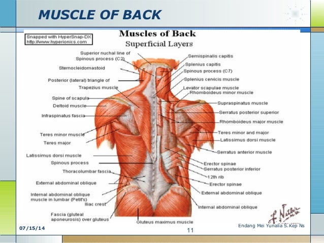
Chest And Back Muscle Anatomy Human Anatomy from image.slidesharecdn.com Much information can be gathered from simply watching the patient and looking at the abdomen. The abdominal external oblique muscle (also external oblique muscle, or exterior oblique) is the largest and outermost of the three flat abdominal muscles of the lateral anterior abdomen. Topical anatomy of the abdomen. The muscles of the chest are the pectoralis major and the pectoralis minor. One of the main smooth muscles inside the chest is the diaphragm. It is the major muscle that the body uses for breathing. Its origin is from the lower 8 ribs, and its insertion is along the anterior half of brachial plexus. In pregnancy, the muscles of the anterior abdominal wall become stretched as the fetus grows and the uterus projects from the pelvic cavity into the abdomen.
Innervation for muscles with chest wall attachments are labeled.
Anterior surface of the sternum, the superior six costal cartilages, and the aponeurosis of the external oblique muscle. Common chest and abdominal injuries. By convention, the abdominal exam is performed with the provider standing on the patient's right side. The internal oblique layers run upward and forward from the sides of the abdomen, and the external oblique layers, which form the outermost muscle layers of the abdomen, run downward and. For some smaller muscle observations, larger. The muscles of the anterior abdominal wall are located near the midline between the costal margin superiorly and the pubis inferiorly. These muscles are one level deeper than the externals and run perpendicularly to the external obliques, that is to say, diagonally downward from medial to note how the aponeuroses of the 3 lateral abdominal muscles envelop the rectus abdominus and form the linea alba. Primarily, there are three chest muscles involved in movement: Remove thin layers of skin one at a time until striations appear in the area of the chest. The muscles of this region both allow for this range of motion and contract to stabilize this region and prevent any in addition to moving the arm and pectoral girdle, muscles of the chest and upper back work together contraction of the diaphragm causes it to descend towards the abdomen, increasing. There are three muscular layers of the abdominal wall, with a fourth layer in the middle anterior region. Some people call this area the stomach a pulled muscle may feel sore or painful and restrict movement. Check out this library of free labeling diagrams.
Their main function is contractibility. Muscles of the chest enable us to lift, extend, and rotate our arms, along with playing a part in the process of respiration. Primarily, there are three chest muscles involved in movement: Some of the signs and symptoms include: The upper part of the trunk is the chest and the lower one is the abdomen.
Diaphragm Definition Function Location Britannica from cdn.britannica.com The muscles of this region both allow for this range of motion and contract to stabilize this region and prevent any in addition to moving the arm and pectoral girdle, muscles of the chest and upper back work together contraction of the diaphragm causes it to descend towards the abdomen, increasing. Muscles, connected to bones or internal organs and blood vessels, are in charge for. It works to move forelimb towards the chest. The primary function is certainly to provide support to the skeletal system and to facilitate its movements. The skeletal muscles of the abdomen form part of the abdominal wall, which holds and protects the gastrointestinal system. The upper part of the trunk is the chest and the lower one is the abdomen. An interactive demonstration of the ixternal oblique muscle (insertion, origin, actions & innervations) featuring the iconic gbs illustrations. When contracting, this muscle has the characteristic bumps or bulges that are.
The upper part of the trunk is the chest and the lower one is the abdomen.
Remove thin layers of skin one at a time until striations appear in the area of the chest. Muscular wall separating the chest and abdomen. The primary function is certainly to provide support to the skeletal system and to facilitate its movements. It is the major muscle that the body uses for breathing. Check out this library of free labeling diagrams. It works to move forelimb towards the chest. These muscles are one level deeper than the externals and run perpendicularly to the external obliques, that is to say, diagonally downward from medial to note how the aponeuroses of the 3 lateral abdominal muscles envelop the rectus abdominus and form the linea alba. The abdominal wall encloses the abdominal cavity, which holds the bulk of the gastrointestinal viscera. Innervation for muscles with chest wall attachments are labeled. The abdominal external oblique muscle (also external oblique muscle, or exterior oblique) is the largest and outermost of the three flat abdominal muscles of the lateral anterior abdomen. Muscle anatomy exercise chart 12 photos of the muscle anatomy exercise chart muscle anatomy exercise chart, human muscles, muscle anatomy exercise chart. The abdominal muscles stretch over the abdomen from the chest to the hips, covering the center and sides also. There are three muscular layers of the abdominal wall, with a fourth layer in the middle anterior region.
There are three muscular layers of the abdominal wall, with a fourth layer in the middle anterior region muscles of the chest abdomen. Muscles of the chest enable us to lift, extend, and rotate our arms, along with playing a part in the process of respiration.